Art & X-rays
- juliet graham
- Apr 26, 2020
- 3 min read
Updated: Apr 30, 2020
Do you love to look up close at artworks? Conservators learn a lot from looking really closely at artworks.

Example of a pastel drawing


The black square in the drawing indicates the area that is magnified


The white circle in the drawing indicates the area shown with 100X magnification
One tool conservators use to look closely and understand more about artworks is an X-ray.
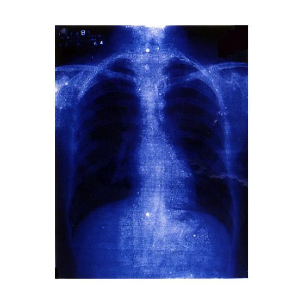
This hand image, below, is one of the first X-rays images ever made. It was made by Willhelm Röntgen on December 22, 1895, and features the left hand of his wife, Anna Bertha Ludwig. It was the first photograph of a human body part using X-rays. When she saw it, she said, "I have seen my death.”

(Photo courtesy of Wikimedia Commons.)
Read more about the discovery of the X-ray at https://www.pbs.org/newshour/health/i-have-seen-my-death-how-the-world-discovered-the-x-ray
Within a year of the discovery of X-rays they had become a fad. People were fascinated by this new way of looking at things. X-ray machines turned up at public exhibitions and lectures, where volunteers from the audience could have their hands or purses X-rayed. People could also buy or build their own X-ray machines at home.

This is an image of an X-ray tram car from 1896. (Courtesy of Wikimedia Commons.)
Read more about the X-ray fad at
https://daily.jstor.org/the-x-ray-craze-of-1896/
The first X-ray of a painting was done in 1896, the year after X-ray was invented. I don’t know when the first painting of an X-ray was made, though! Here is a print that looks a little like an X-ray by artist Les Levine. This print is from the University of Lethbridge art collection, and was made almost 70 years after X-rays were invented.
Title: Wellesley Hospital (Toronto 20 Suite), 1964. By Les Levine From the University of Lethbridge art collection; gift of Avrom Isaacs, Toronto, 1986
A painting that has gotten more than its fair share of X-rays is “Girl with a Pearl Earring” by Vermeer, owned by the Mauritshuis Museum in Holland. Its first X-ray was taken in the 1920s by Alan Burroughs from the Fogg Museum at Harvard University.


The full colour image is a recent photograph of "Girl with a Pearl Earring" by Johannes Vermeer, painted circa 1665, and now owned by the Mauritshaus Museum in the Hague. The black and white image is a circa 1920 X-ray of "Girl with a Pearl Earring". The X-ray image is owned by the Straus Centre for Conservation and Technical Studies at the Harvard Art Museums, in the Alan Burroughs collection of X-rays.
Alan Burroughs was an art conservator who wanted to use science to learn more about art. He did a systematic study of many paintings from many different countries; He would usually take images of characteristic details from at least four paintings by a significant artist, and use them to find out relationships between paintings by the same artist, or to help to discover who a painting was created by if it was unsigned or had any doubts surrounding it regarding its authorship.
If you want to see more, these X-ray documentation photos have been digitized and can be viewed here: https://www.harvardartmuseums.org/publications/special-collections/alan-burroughs-collection-of-x-radiographs?group=Alan+Burroughs+Collection+of+X-Radiographs
Look at this X-ray below and how it shows a different composition hidden underneath the finished painting. You can see a regular photo of the painting on the National Gallery London website and find out which version was the final one, and which one got painted over. https://www.nationalgallery.org.uk/paintings/francisco-de-goya-dona-isabel-de-porcel (And, how nice! It is one of those high-res images that you can look really closely at!)

"Portrait of Isobel de Porcel" by Francisco Goya. (X-ray image from Wikimedia Commons.)

"Girl with a Pearl Earring" by Johannes Vermeer, circa 1665. This is how it looks installed at the Mauritshaus Museum in the Hague.
2 years ago, in 2018, “Girl with a Pearl Earring” got an incredibly thorough examination, which is documented in the Mauritshuis website in an entertaining and informative 20 part illustrated story called, “Girl with a Blog”. In it you will learn from Abbie Vandivere, the lead conservator, about the high-tech scientific examination techniques used to find out things like:
-the painting’s history: for example, how did it get to be so dirty and in such poor condition that it was hardly recognizable and sold at auction in 1881 for less than 50 dollars (translated into today’s money)
- Why does the X-ray show areas of loss but in regular lighting the painting looks just fine?
-Did Vermeer paint the pearl earring with one highlight…or two highlights? And is it really a pearl?
-How did artists make paint around 1665 when Vermeer was working? And why would they store paint in an animal bladder?
-Watch cool videos about how to do your makeup to look just like the girl, and how to turn blue rocks into blue paint! - And learn how two weeks of round-the-clock scientific examination in 2018 has made the Girl with the Pearl Earring one of the most thoroughly researched paintings in the world.
Find the blog here: https://www.mauritshuis.nl/en/explore/restoration-and-research/girl-with-a-blog/


This fascinating article beautifully captures the historic origins of X-ray imaging, tracing back to Willhelm Röntgen’s revolutionary work and the profound emotional reaction it evoked—underscoring how radiographic technology forever transformed medicine, conservation, and diagnostics.
Just as X-rays revealed hidden structures, a White Screen helps uncover screen flaws or dust on devices like Dell, HP, iPhones, and Smart TVs.
Try Bright WhiteScreen on any browser for OLED/QLED testing, device cleaning, or room lighting—essential for professionals, artists, and curious minds alike.